225
views
0
recommends
+1 Recommend
0 collections
    0
    shares
      • Record: found
      • Abstract: found
      • Article: not found

      Modeling the mitochondrial cardiomyopathy of Barth syndrome with iPSC and heart-on-chip technologies

      research-article

      Read this article at

      ScienceOpenPublisherPMC
          There is no author summary for this article yet. Authors can add summaries to their articles on ScienceOpen to make them more accessible to a non-specialist audience.

          Abstract

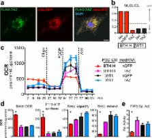
          Studying monogenic mitochondrial cardiomyopathies may yield insights into mitochondrial roles in cardiac development and disease. Here, we combine patient-derived and genetically engineered iPSCs with tissue engineering to elucidate the pathophysiology underlying the cardiomyopathy of Barth syndrome (BTHS), a mitochondrial disorder caused by mutation of the gene Tafazzin (TAZ). Using BTHS iPSC-derived cardiomyocytes (iPSC-CMs), we defined metabolic, structural, and functional abnormalities associated with TAZ mutation. BTHS iPSC-CMs assembled sparse and irregular sarcomeres, and engineered BTHS “heart on chip” tissues contracted weakly. Gene replacement and genome editing demonstrated that TAZ mutation is necessary and sufficient for these phenotypes. Sarcomere assembly and myocardial contraction abnormalities occurred in the context of normal whole cell ATP levels. Excess levels of reactive oxygen species mechanistically linked TAZ mutation to impaired cardiomyocyte function. Our study provides new insights into the pathogenesis of Barth syndrome, suggests new treatment strategies, and advances iPSC-based in vitro modeling of cardiomyopathy.

          Related collections

          Most cited references28

          • Record: found
          • Abstract: not found
          • Article: not found

          The Tension of Metallic Films Deposited by Electrolysis

            Bookmark
            • Record: found
            • Abstract: found
            • Article: not found

            hESC-Derived Cardiomyocytes Electrically Couple and Suppress Arrhythmias in Injured Hearts

            Transplantation studies in mice and rats have shown that human embryonic stem cell-derived cardiomyocytes (hESC-CMs) can improve the function of infarcted hearts 1–3 , but two critical issues related to their electrophysiological behavior in vivo remain unresolved. First, the risk of arrhythmias following hESC-CM transplantation in injured hearts has not been determined. Second, the electromechanical integration of hESC-CMs in injured hearts has not been demonstrated, so it is unclear if these cells improve contractile function directly through addition of new force-generating units. Here we use a guinea pig model to show hESC-CM grafts in injured hearts protect against arrhythmias and can contract synchronously with host muscle. Injured hearts with hESC-CM grafts show improved mechanical function and a significantly reduced incidence of both spontaneous and induced ventricular tachycardia (VT). To assess the activity of hESC-CM grafts in vivo, we transplanted hESC-CMs expressing the genetically-encoded calcium sensor, GCaMP3 4, 5 . By correlating the GCaMP3 fluorescent signal with the host ECG, we found that grafts in uninjured hearts have consistent 1:1 host-graft coupling. Grafts in injured hearts are more heterogeneous and typically include both coupled and uncoupled regions. Thus, human myocardial grafts meet physiological criteria for true heart regeneration, providing support for the continued development of hESC-based cardiac therapies for both mechanical and electrical repair.
              Bookmark
              • Record: found
              • Abstract: found
              • Article: not found

              Patient-specific induced pluripotent stem cells as a model for familial dilated cardiomyopathy.

              Characterized by ventricular dilatation, systolic dysfunction, and progressive heart failure, dilated cardiomyopathy (DCM) is the most common form of cardiomyopathy in patients. DCM is the most common diagnosis leading to heart transplantation and places a significant burden on healthcare worldwide. The advent of induced pluripotent stem cells (iPSCs) offers an exceptional opportunity for creating disease-specific cellular models, investigating underlying mechanisms, and optimizing therapy. Here, we generated cardiomyocytes from iPSCs derived from patients in a DCM family carrying a point mutation (R173W) in the gene encoding sarcomeric protein cardiac troponin T. Compared to control healthy individuals in the same family cohort, cardiomyocytes derived from iPSCs from DCM patients exhibited altered regulation of calcium ion (Ca(2+)), decreased contractility, and abnormal distribution of sarcomeric α-actinin. When stimulated with a β-adrenergic agonist, DCM iPSC-derived cardiomyocytes showed characteristics of cellular stress such as reduced beating rates, compromised contraction, and a greater number of cells with abnormal sarcomeric α-actinin distribution. Treatment with β-adrenergic blockers or overexpression of sarcoplasmic reticulum Ca(2+) adenosine triphosphatase (Serca2a) improved the function of iPSC-derived cardiomyocytes from DCM patients. Thus, iPSC-derived cardiomyocytes from DCM patients recapitulate to some extent the morphological and functional phenotypes of DCM and may serve as a useful platform for exploring disease mechanisms and for drug screening.
                Bookmark

                Author and article information

                Journal
                9502015
                8791
                Nat Med
                Nat. Med.
                Nature medicine
                1078-8956
                1546-170X
                8 August 2014
                11 May 2014
                June 2014
                01 December 2014
                : 20
                : 6
                : 616-623
                Affiliations
                [1 ]Department of Cardiology, Boston Children’s Hospital, Boston, MA, 02115, USA
                [2 ]Wyss Institute for Biologically Inspired Engineering, School of Engineering and Applied Sciences, Harvard University, Cambridge, MA, 02138, USA
                [3 ]Department of Genetics, Harvard Medical School, Boston, MA, 02115, USA
                [4 ]Boston Children’s Hospital Department of Medicine, Division of Genetics, Boston Children’s Hospital, Boston, MA, 02115, USA
                [5 ]Allele Biotechnology & Pharmaceuticals, Inc., San Diego, CA, 92121, USA
                [6 ]Department of Clinical Chemistry and Pediatrics, Laboratory Genetic Metabolic Disease, Academic Medical Center, Amsterdam, 1105 AZ, The Netherlands
                [7 ]Department of Pathology, Center for Cardiovascular Biology and Institute for Stem Cell and Regenerative Medicine, University of Washington, Seattle, WA, 98109, USA
                [8 ]Departments of Bioengineering and Medicine/Cardiology, University of Washington, Seattle, WA, 98109, USA
                [9 ]Department of Cell and Molecular Biology and Medicine, Karolinska Institutet, 17177, Stockholm, Sweden
                [10 ]Division of Metabolism, Kennedy Krieger Institute, Baltimore, MD, 21205, USA
                [11 ]Harvard Stem Cell Institute, Harvard University, Cambridge, MA, 02138, USA
                Author notes
                [¥ ]Correspondence to: W.T.P. 300 Longwood Ave, Boston, MA 02115, USA. wpu@ 123456enders.tch.harvard.edu . Phone: 617-919-2091. Fax: 617-730-0140. K.K.P. 29 Oxford Street, Pierce Hall Cambridge, MA 02130, USA. kkparker@ 123456seas.harvard.edu . Phone: 617-495-2850. Fax: 617-496-1793
                [*]

                contributed equally

                Article
                NIHMS579142
                10.1038/nm.3545
                4172922
                24813252
                aa4af9b8-dcf1-4791-ab96-e2720a0c4127
                History
                Categories
                Article

                Medicine
                Medicine

                Comments

                Comment on this article