1
views
0
recommends
+1 Recommend
0 collections
    0
    shares
      • Record: found
      • Abstract: found
      • Article: found
      Is Open Access

      Short-term acute bright light exposure induces a prolonged anxiogenic effect in mice via a retinal ipRGC-CeA circuit

      research-article

      Read this article at

      Bookmark
          There is no author summary for this article yet. Authors can add summaries to their articles on ScienceOpen to make them more accessible to a non-specialist audience.

          Abstract

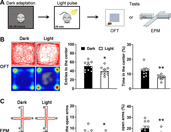
          Light modulates mood through various retina-brain pathways. We showed that mice treated with short-term acute bright light exposure displayed anxiety-related phenotypes in a prolonged manner even after the termination of the exposure. Such a postexposure anxiogenic effect depended upon melanopsin-based intrinsically photosensitive retinal ganglion cell (ipRGC) activities rather than rod/cone photoreceptor inputs. Chemogenetic manipulation of specific central nuclei demonstrated that the ipRGC–central amygdala (CeA) visual circuit played a key role in this effect. The corticosterone system was likely to be involved in this effect, as evidenced by enhanced expression of the glucocorticoid receptor (GR) protein in the CeA and the bed nucleus of the stria terminalis and by the absence of this effect in animals treated with the GR antagonist. Together, our findings reveal a non-image forming visual circuit specifically designed for “the delayed” extinction of anxiety against potential threats, thus conferring a survival advantage.

          Abstract

          Abstract

          Intrinsically photosensitive ganglion cells in the retina activated by bright light are involved in anxiety.

          Related collections

          Most cited references77

          • Record: found
          • Abstract: found
          • Article: not found

          Phototransduction by retinal ganglion cells that set the circadian clock.

          Light synchronizes mammalian circadian rhythms with environmental time by modulating retinal input to the circadian pacemaker-the suprachiasmatic nucleus (SCN) of the hypothalamus. Such photic entrainment requires neither rods nor cones, the only known retinal photoreceptors. Here, we show that retinal ganglion cells innervating the SCN are intrinsically photosensitive. Unlike other ganglion cells, they depolarized in response to light even when all synaptic input from rods and cones was blocked. The sensitivity, spectral tuning, and slow kinetics of this light response matched those of the photic entrainment mechanism, suggesting that these ganglion cells may be the primary photoreceptors for this system.
            Bookmark
            • Record: found
            • Abstract: found
            • Article: not found

            Melanopsin-expressing ganglion cells in primate retina signal colour and irradiance and project to the LGN.

            Human vision starts with the activation of rod photoreceptors in dim light and short (S)-, medium (M)-, and long (L)- wavelength-sensitive cone photoreceptors in daylight. Recently a parallel, non-rod, non-cone photoreceptive pathway, arising from a population of retinal ganglion cells, was discovered in nocturnal rodents. These ganglion cells express the putative photopigment melanopsin and by signalling gross changes in light intensity serve the subconscious, 'non-image-forming' functions of circadian photoentrainment and pupil constriction. Here we show an anatomically distinct population of 'giant', melanopsin-expressing ganglion cells in the primate retina that, in addition to being intrinsically photosensitive, are strongly activated by rods and cones, and display a rare, S-Off, (L + M)-On type of colour-opponent receptive field. The intrinsic, rod and (L + M) cone-derived light responses combine in these giant cells to signal irradiance over the full dynamic range of human vision. In accordance with cone-based colour opponency, the giant cells project to the lateral geniculate nucleus, the thalamic relay to primary visual cortex. Thus, in the diurnal trichromatic primate, 'non-image-forming' and conventional 'image-forming' retinal pathways are merged, and the melanopsin-based signal might contribute to conscious visual perception.
              Bookmark
              • Record: found
              • Abstract: not found
              • Article: not found

              The role of the amygdala in fear and anxiety.

              M DAVIS (1992)
                Bookmark

                Author and article information

                Contributors
                Role: ConceptualizationRole: Formal analysisRole: InvestigationRole: MethodologyRole: Project administrationRole: SoftwareRole: ValidationRole: VisualizationRole: Writing - original draftRole: Writing - review & editing
                Role: Formal analysisRole: InvestigationRole: Methodology
                Role: InvestigationRole: Writing - review & editing
                Role: InvestigationRole: Resources
                Role: Formal analysisRole: InvestigationRole: Validation
                Role: InvestigationRole: MethodologyRole: ResourcesRole: ValidationRole: Writing - review & editing
                Role: Investigation
                Role: ConceptualizationRole: Data curationRole: Formal analysisRole: Funding acquisitionRole: InvestigationRole: MethodologyRole: Project administrationRole: ResourcesRole: SupervisionRole: ValidationRole: VisualizationRole: Writing - original draftRole: Writing - review & editing
                Role: Formal analysisRole: Investigation
                Role: Formal analysis
                Role: Formal analysisRole: Methodology
                Role: Formal analysisRole: Funding acquisitionRole: Project administrationRole: VisualizationRole: Writing - review & editing
                Role: ConceptualizationRole: Formal analysisRole: Funding acquisitionRole: Project administrationRole: ValidationRole: Writing - original draftRole: Writing - review & editing
                Role: ConceptualizationRole: Funding acquisitionRole: SupervisionRole: ValidationRole: Writing - review & editing
                Journal
                Sci Adv
                Sci Adv
                sciadv
                advances
                Science Advances
                American Association for the Advancement of Science
                2375-2548
                March 2023
                22 March 2023
                : 9
                : 12
                : eadf4651
                Affiliations
                [ 1 ]State Key Laboratory of Medical Neurobiology and MOE Frontiers Center for Brain Science, Institutes of Brain Science, Fudan University, Shanghai, China.
                [ 2 ]Department of Ophthalmology, Shanghai General Hospital, National Clinical Research Center for Eye Diseases, Shanghai Key Laboratory of Ocular Fundus Diseases, Shanghai Engineering Center for Visual Science and Photomedicine, Shanghai Engineering Center for Precise Diagnosis and Treatment of Eye Diseases, Shanghai, China.
                Author notes
                [* ]Corresponding author. Email: xlyang@ 123456fudan.edu.cn (X.-L.Y.); sjweng@ 123456fudan.edu.cn (S.-J.W.)
                Author information
                https://orcid.org/0000-0002-7198-134X
                https://orcid.org/0000-0003-3647-6310
                https://orcid.org/0000-0001-8047-6751
                https://orcid.org/0000-0001-5048-3379
                https://orcid.org/0000-0001-6623-9629
                https://orcid.org/0000-0002-3382-2655
                https://orcid.org/0000-0001-6550-892X
                https://orcid.org/0000-0003-2128-9950
                Article
                adf4651
                10.1126/sciadv.adf4651
                10032603
                36947616
                f3a0121c-c8a6-4e77-87b7-8948f3b46583
                Copyright © 2023 The Authors, some rights reserved; exclusive licensee American Association for the Advancement of Science. No claim to original U.S. Government Works. Distributed under a Creative Commons Attribution NonCommercial License 4.0 (CC BY-NC).

                This is an open-access article distributed under the terms of the Creative Commons Attribution-NonCommercial license, which permits use, distribution, and reproduction in any medium, so long as the resultant use is not for commercial advantage and provided the original work is properly cited.

                History
                : 29 October 2022
                : 17 February 2023
                Funding
                Funded by: FundRef http://dx.doi.org/10.13039/501100001809, National Natural Science Foundation of China;
                Award ID: 81790640
                Funded by: FundRef http://dx.doi.org/10.13039/501100001809, National Natural Science Foundation of China;
                Award ID: 82070993
                Funded by: FundRef http://dx.doi.org/10.13039/501100001809, National Natural Science Foundation of China;
                Award ID: 31571072
                Funded by: FundRef http://dx.doi.org/10.13039/501100001809, National Natural Science Foundation of China;
                Award ID: 32070989
                Funded by: FundRef http://dx.doi.org/10.13039/501100001809, National Natural Science Foundation of China;
                Award ID: 31872766
                Funded by: FundRef http://dx.doi.org/10.13039/501100001809, National Natural Science Foundation of China;
                Award ID: 31571075
                Funded by: Ministry of Science and Technology, Government of the People’s Republic of China;
                Award ID: 2022ZD0208604
                Funded by: Ministry of Science and Technology, Government of the People’s Republic of China;
                Award ID: 2022ZD0208605
                Funded by: Shanghai Municipal Science and Technology Major Project;
                Award ID: 2018SHZDZX01
                Funded by: Shanghai Center for Brain Science and Brain-Inspired Technology;
                Award ID: N.A.
                Funded by: FundRef http://dx.doi.org/10.13039/501100012151, Sanming Project of Medicine in Shenzhen;
                Award ID: SZSM202011015
                Funded by: ZJLab;
                Award ID: N.A.
                Categories
                Research Article
                Neuroscience
                SciAdv r-articles
                Life Sciences
                Neuroscience
                Neuroscience
                Custom metadata
                Anne Suarez

                Comments

                Comment on this article