2
views
0
recommends
+1 Recommend
0 collections
    0
    shares
      • Record: found
      • Abstract: found
      • Article: found
      Is Open Access

      Scalp‐recorded N40 visual evoked potential: Sensory and attentional properties

      research-article

      Read this article at

      Bookmark
          There is no author summary for this article yet. Authors can add summaries to their articles on ScienceOpen to make them more accessible to a non-specialist audience.

          Abstract

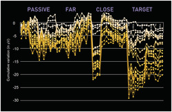
          N40 is a well‐known component of evoked potentials with respect to the auditory and somatosensory modality but not much recognized with regard to the visual modality. To be detected with event‐related potentials (ERPs), it requires an optimal signal‐to‐noise ratio. To investigate the nature of visual N40, we recorded EEG/ERP signals from 20 participants. Each of them was presented with 1800 spatial frequency gratings of 0.75, 1.5, 3 and 6 c/deg. Data were collected from 128 sites while participants were engaged in both passive viewing and attention conditions. N40 (30–55 ms) was modulated by alertness and selective attention; in fact, it was larger to targets than irrelevant and passively viewed spatial frequency gratings. Its strongest intracranial sources were the bilateral thalamic nuclei of pulvinar, according to swLORETA. The active network included precuneus, insula and inferior parietal lobule. An N80 component (60–90 ms) was also identified, which was larger to targets than irrelevant/passive stimuli and more negative to high than low spatial frequencies. In contrast, N40 was not sensitive to spatial frequency per se, nor did it show a polarity inversion as a function of spatial frequency. Attention, alertness and spatial frequency effects were also found for the later components P1, N2 and P300. The attentional effects increased in magnitude over time. The data showed that ERPs can pick up the earliest synchronized activity, deriving in part from thalamic nuclei, before the visual information has actually reached the occipital cortex.

          Abstract

          Recorded for the first time on the human scalp electrical signals deriving from the thalamus 40 ms after the onset of visual stimulation. The EEG data showed that visual evoked potentials (VEPs) can detect signals that originate from subcortical structures before visual information has actually reached the occipital cortex. These findings indicate that the thalamus not only transmits sensory signals but also acts as an attentional filter. This paper describes sensory and attentional properties of newly defined N40 component of VEPs.

          Related collections

          Most cited references94

          • Record: found
          • Abstract: found
          • Article: not found

          Updating P300: an integrative theory of P3a and P3b.

          The empirical and theoretical development of the P300 event-related brain potential (ERP) is reviewed by considering factors that contribute to its amplitude, latency, and general characteristics. The neuropsychological origins of the P3a and P3b subcomponents are detailed, and how target/standard discrimination difficulty modulates scalp topography is discussed. The neural loci of P3a and P3b generation are outlined, and a cognitive model is proffered: P3a originates from stimulus-driven frontal attention mechanisms during task processing, whereas P3b originates from temporal-parietal activity associated with attention and appears related to subsequent memory processing. Neurotransmitter actions associating P3a to frontal/dopaminergic and P3b to parietal/norepinephrine pathways are highlighted. Neuroinhibition is suggested as an overarching theoretical mechanism for P300, which is elicited when stimulus detection engages memory operations.
            Bookmark
            • Record: found
            • Abstract: found
            • Article: not found

            Low resolution electromagnetic tomography: a new method for localizing electrical activity in the brain.

            This paper presents a new method for localizing the electric activity in the brain based on multichannel surface EEG recordings. In contrast to the models presented up to now the new method does not assume a limited number of dipolar point sources nor a distribution on a given known surface, but directly computes a current distribution throughout the full brain volume. In order to find a unique solution for the 3-dimensional distribution among the infinite set of different possible solutions, the method assumes that neighboring neurons are simultaneously and synchronously activated. The basic assumption rests on evidence from single cell recordings in the brain that demonstrates strong synchronization of adjacent neurons. In view of this physiological consideration the computational task is to select the smoothest of all possible 3-dimensional current distributions, a task that is a common procedure in generalized signal processing. The result is a true 3-dimensional tomography with the characteristic that localization is preserved with a certain amount of dispersion, i.e., it has a relatively low spatial resolution. The new method, which we call Low Resolution Electromagnetic Tomography (LORETA) is illustrated with two different sets of evoked potential data, the first showing the tomography of the P100 component to checkerboard stimulation of the left, right, upper and lower hemiretina, and the second showing the results for the auditory N100 component and the two cognitive components CNV and P300. A direct comparison of the tomography results with those obtained from fitting one and two dipoles illustrates that the new method provides physiologically meaningful results while dipolar solutions fail in many situations. In the case of the cognitive components, the method offers new hypotheses on the location of higher cognitive functions in the brain.
              Bookmark
              • Record: found
              • Abstract: found
              • Article: not found

              Electrophysiological correlates of feature analysis during visual search.

              Event-related brain potentials (ERPs) were recorded from normal young adults during visual search tasks in which the stimulus arrays contained either eight identical items (homogeneous arrays) or seven identical items and one deviant item (pop-out arrays). Four experiments were conducted in which different classes of stimulus arrays were designated targets and the remaining stimulus arrays were designated nontargets. In Experiments 1 and 2, both target and nontarget pop-out stimuli elicited an enhanced anterior N2 wave and a contralaterally larger posterior P1 wave, but Experiments 3 and 4 demonstrated that these components do not reflect fully automatic pop-out detection processes. In all four experiments, target pop-outs elicited enlarged anterior P2, posterior N2, occipital P3, and parietal P3 waves. The target-elicited posterior N2 wave contained a contralateral subcomponent (N2pc) that exhibited a focus over occipital cortex in maps of current source density. The overall pattern of results was consistent with guided search models in which preattentive stimulus information is used to guide attention to task-relevant stimuli.
                Bookmark

                Author and article information

                Contributors
                mado.proverbio@unimib.it
                Journal
                Eur J Neurosci
                Eur J Neurosci
                10.1111/(ISSN)1460-9568
                EJN
                The European Journal of Neuroscience
                John Wiley and Sons Inc. (Hoboken )
                0953-816X
                1460-9568
                27 September 2021
                October 2021
                : 54
                : 7 ( doiID: 10.1111/ejn.v54.7 )
                : 6553-6574
                Affiliations
                [ 1 ] Department of Psychology University of Milano‐Bicocca Milan Italy
                [ 2 ] Milan Center for Neuroscience (NeuroMi) University of Milano‐Bicocca Milan Italy
                [ 3 ] School of Psychology Vita Salute San Raffaele University Milan Italy
                Author notes
                [*] [* ] Correspondence

                Alice Mado Proverbio, Department of Psychology, University of Milano‐Bicocca, Piazza dell'Ateneo Nuovo 1, Milan, Italy.

                Email: mado.proverbio@ 123456unimib.it

                Author information
                https://orcid.org/0000-0002-5138-1523
                Article
                EJN15443
                10.1111/ejn.15443
                9293152
                34486754
                deed9e55-26b2-4d05-9e03-e985e5d38c2b
                © 2021 The Authors. European Journal of Neuroscience published by Federation of European Neuroscience Societies and John Wiley & Sons Ltd.

                This is an open access article under the terms of the http://creativecommons.org/licenses/by/4.0/ License, which permits use, distribution and reproduction in any medium, provided the original work is properly cited.

                History
                : 23 August 2021
                : 30 November 2020
                : 28 August 2021
                Page count
                Figures: 13, Tables: 2, Pages: 22, Words: 13568
                Categories
                Research Report
                Systems Neuroscience
                Custom metadata
                2.0
                October 2021
                Converter:WILEY_ML3GV2_TO_JATSPMC version:6.1.7 mode:remove_FC converted:18.07.2022

                Neurosciences
                attention,eeg/erps,gratings,lgn,spatial frequency,thalamus,vep
                Neurosciences
                attention, eeg/erps, gratings, lgn, spatial frequency, thalamus, vep

                Comments

                Comment on this article