0
views
0
recommends
+1 Recommend
0 collections
    0
    shares
      • Record: found
      • Abstract: found
      • Article: found
      Is Open Access

      Randomized comparison of oblique and perpendicular stabilizers for minimally invasive repair of pectus excavatum

      research-article

      Read this article at

      Bookmark
          There is no author summary for this article yet. Authors can add summaries to their articles on ScienceOpen to make them more accessible to a non-specialist audience.

          Abstract

          OBJECTIVES

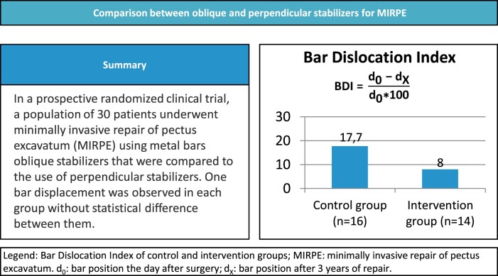
          Bar dislocation is one of the most feared complications of the minimally invasive repair of pectus excavatum.

          METHODS

          Prospective randomized parallel-group clinical trial intending to assess whether oblique stabilizers can reduce bar displacement in comparison with regular stabilizers used in minimally invasive repair of pectus excavatum. Additionally, we evaluated pain, quality of life and other postoperative complications. Participants were randomly assigned to surgery with perpendicular ( n = 16) or oblique stabilizers ( n = 14) between October 2017 and September 2018 and followed for 3 years. Bar displacements were evaluated with the bar displacement index. Pain scores were evaluated through visual analogue scale and quality of life through the Pectus Excavatum Evaluation Questionnaire.

          RESULTS

          Control group average displacement index was 17.7 (±26.7) and intervention group average displacement index was 8.2 (±10.9). There was 1 reoperation in each group that required correction with 2 bars. Bar displacement was similar among groups ( P = 0.12). No other complications were recorded. There was no statistically significant difference on pain score. There was a significant difference between pre- and postoperative composite scores of the participants’ body image domain and psycho-social aspects in both groups. The difference between the pre- and postoperative participants’ perception of physical difficulties was greater and statistically significant in the intervention group.

          CONCLUSIONS

          There was no statistical difference in the use of perpendicular or oblique stabilizers, but the availability of different models of stabilizers during the study suggested that this can be advantageous. The trial is registered at ClinicalTrials.gov, number NCT03087734.

          Abstract

          Pectus excavatum (PE) is associated with shortness of breath, exercise intolerance, low self-esteem and depression [1–3] and to date there is no effective non-invasive treatment [4].

          Related collections

          Most cited references29

          • Record: found
          • Abstract: found
          • Article: not found
          Is Open Access

          CONSORT 2010 Statement: updated guidelines for reporting parallel group randomised trials

          The CONSORT statement is used worldwide to improve the reporting of randomised controlled trials. Kenneth Schulz and colleagues describe the latest version, CONSORT 2010, which updates the reporting guideline based on new methodological evidence and accumulating experience
            Bookmark
            • Record: found
            • Abstract: found
            • Article: not found

            Experience and modification update for the minimally invasive Nuss technique for pectus excavatum repair in 303 patients.

            The aim of this study is to review the new technical modifications and results of 303 patients who have had pectus excavatum repair utilizing the minimally invasive technique. A retrospective chart review was conducted of 303 patients undergoing minimally invasive pectus repair from 1987 through August 2000. Since 1997, a standardized treatment pathway was implemented, and 261 of the 303 patients have been treated on this pathway. Preoperative evaluation included computed tomography (CT) scan, pulmonary function tests (PFT), and cardiac evaluations with electrocardiogram (EKG) and echocardiogram. Indications for operation included at least 2 of the following: progression of the deformity, exercise intolerance or restrictive disease on PFT, Haller CT index greater than 3.2, mitral valve prolapse (MVP), or cardiac compression. Technical and design modifications since 1998 have included routine thoracoscopy, the use of an introducer/dissector for creating the substernal tunnel and elevating the sternum, and routine use of a wired lateral stabilizer to prevent bar displacement. The bar is removed as an outpatient procedure in 2 to 4 years. In 303 patients undergoing minimally invasive pectus repairs, single bars were used in 87% and double in 13%. Lateral stabilizers were applied in 70% of patients and were wired for further stability in 65%. Bar shifts before the use of stabilizers were 15%, which decreased to 6% after stabilizers were placed and 5% with a wired stabilizer. Excellent results were noted in 85% with failure in only 1 patient. Complications included pneumothorax with spontaneous resolution in half of the patients and pericarditis in 7. The minimally invasive technique has evolved into an effective method of pectus excavatum repair. Modifications of the technique have reduced complications. Long-term results continue to be excellent. Copyright 2002 by W.B. Saunders Company.
              Bookmark
              • Record: found
              • Abstract: found
              • Article: not found

              Surgical repair of pectus excavatum markedly improves body image and perceived ability for physical activity: multicenter study.

              This study evaluated changes in both physical and psychosocial quality of life reported by the parent and child after surgical repair of pectus excavatum. As part of a multicenter study of pectus excavatum, a previously validated tool called the Pectus Excavatum Evaluation Questionnaire was administered by the research coordinator, via telephone, to parents and patients (8-21 years of age) before and 1 year after surgery. Eleven North American children's hospitals participated. From 2001 to 2006, 264 patients and 291 parents completed the initial questionnaire, and 247 patients and 274 parents completed the postoperative questionnaire. Responses used a Likert-type scale of 1 to 4, reflecting the extent or frequency of a particular experience, with higher values conveying less-desirable experience. Preoperative psychosocial functioning was unrelated to objective pectus excavatum severity (computed tomographic index). Patients and their parents reported significant positive postoperative changes. Improvements occurred in both physical and psychosocial functioning, including less social self-consciousness and a more-favorable body image. For children, the body image component improved from 2.30+/-0.62 (mean+/-SD) to 1.40+/-0.42 after surgery and the physical difficulties component improved from 2.11+/-0.82 to 1.37+/-0.44. For the parent questionnaire, the child's emotional difficulties improved from 1.81+/-0.70 to 1.24+/-0.36, social self-consciousness improved from 2.86+/-1.03 to 1.33+/-0.68, and physical difficulties improved from 2.14+/-0.75 to 1.32+/-0.39. Ninety-seven percent of patients thought that surgery improved how their chest looked. Surgical repair of pectus excavatum can significantly improve the body image difficulties and limitations on physical activity experienced by patients. These results should prompt physicians to consider the physiologic and psychological implications of pectus excavatum just as they would any other physical deformity known to have such consequences.
                Bookmark

                Author and article information

                Contributors
                Journal
                Interdiscip Cardiovasc Thorac Surg
                Interdiscip Cardiovasc Thorac Surg
                icvts
                Interdisciplinary Cardiovascular and Thoracic Surgery
                Oxford University Press
                2753-670X
                March 2024
                16 March 2024
                16 March 2024
                : 38
                : 3
                : ivae040
                Affiliations
                Divisao Cirurgia Toracica, Instituto do Coracao, Hospital das Clinicas HCFMUSP, Faculdade de Medicina, Universidade de Sao Paulo, Sao Paulo, SP, Brazil
                Thoracic Surgery Division, Hospital Infantil Sabará , São Paulo, Brazil
                Divisao Cirurgia Toracica, Instituto do Coracao, Hospital das Clinicas HCFMUSP, Faculdade de Medicina, Universidade de Sao Paulo, Sao Paulo, SP, Brazil
                Divisao Cirurgia Toracica, Instituto do Coracao, Hospital das Clinicas HCFMUSP, Faculdade de Medicina, Universidade de Sao Paulo, Sao Paulo, SP, Brazil
                Divisao Cirurgia Toracica, Instituto do Coracao, Hospital das Clinicas HCFMUSP, Faculdade de Medicina, Universidade de Sao Paulo, Sao Paulo, SP, Brazil
                Divisao Cirurgia Toracica, Instituto do Coracao, Hospital das Clinicas HCFMUSP, Faculdade de Medicina, Universidade de Sao Paulo, Sao Paulo, SP, Brazil
                Divisao Cirurgia Toracica, Instituto do Coracao, Hospital das Clinicas HCFMUSP, Faculdade de Medicina, Universidade de Sao Paulo, Sao Paulo, SP, Brazil
                Divisao Cirurgia Toracica, Instituto do Coracao, Hospital das Clinicas HCFMUSP, Faculdade de Medicina, Universidade de Sao Paulo, Sao Paulo, SP, Brazil
                Divisao Cirurgia Toracica, Instituto do Coracao, Hospital das Clinicas HCFMUSP, Faculdade de Medicina, Universidade de Sao Paulo, Sao Paulo, SP, Brazil
                Divisao Cirurgia Toracica, Instituto do Coracao, Hospital das Clinicas HCFMUSP, Faculdade de Medicina, Universidade de Sao Paulo, Sao Paulo, SP, Brazil
                Divisao Cirurgia Toracica, Instituto do Coracao, Hospital das Clinicas HCFMUSP, Faculdade de Medicina, Universidade de Sao Paulo, Sao Paulo, SP, Brazil
                Author notes
                Corresponding author. Thoracic Surgery Division, Heart Institute (InCor), University of Sao Paulo Medical School, R. Itambe, 367, ap 151A, SP 01239-001, Brazil. Tel: +55-11-99653-5030; e-mail: tedde@ 123456usp.br (M.L. Tedde).
                Author information
                https://orcid.org/0000-0002-8178-4243
                https://orcid.org/0000-0001-5956-2962
                https://orcid.org/0000-0002-2385-7707
                https://orcid.org/0000-0002-8979-5035
                https://orcid.org/0000-0002-1226-085X
                https://orcid.org/0000-0002-9908-2215
                https://orcid.org/0000-0001-7243-5343
                Article
                ivae040
                10.1093/icvts/ivae040
                10973937
                38492558
                dee8dcd3-f0c2-42d4-88ee-e87bc023e92a
                © The Author(s) 2024. Published by Oxford University Press on behalf of the European Association for Cardio-Thoracic Surgery.

                This is an Open Access article distributed under the terms of the Creative Commons Attribution License ( https://creativecommons.org/licenses/by/4.0/), which permits unrestricted reuse, distribution, and reproduction in any medium, provided the original work is properly cited.

                History
                : 30 November 2023
                : 22 January 2024
                : 14 March 2024
                : 27 March 2024
                Page count
                Pages: 8
                Funding
                Funded by: Fundação de Amparo à Pesquisa do Estado de São Paulo, DOI 10.13039/501100001807;
                Funded by: Traumec Tecnologia e Implantes Ltda;
                Categories
                Thoracic Non-oncology
                Original Article
                Eacts/147
                Eacts/154
                AcademicSubjects/MED00920

                funnel chest,chest wall,minimally invasive surgery,clinical trials,protocol

                Comments

                Comment on this article