14
views
0
recommends
+1 Recommend
0 collections
    0
    shares
      • Record: found
      • Abstract: found
      • Article: found
      Is Open Access

      Surgical spectral imaging

      research-article
      a , b , * , a , c , d , e , f , a , c
      Medical Image Analysis
      Elsevier
      Multispectral imaging, Hyperspectral imaging, Minimally-invasive surgery, Computational imaging, AI, Artificial intelligence, AOTF, Acousto-optic tuneable filter, CNN, Convolutional neural network, CT, Computed tomography, DMD, Digital micromirror device, DPF, Differential pathlength factor, EMCCD, Electron-multiplying charge-coupled device, FIGS, Fluorescence image-guided surgery, FWHM, Full-width at half-maximum, GI, Gastrointestinal, HSI, Hyperspectral imaging, INN, Invertible neural network, LCTF, Liquid crystal tuneable filter, LED, Light emitting diode, LOOCV, Leave-one-out cross validation, MIS, Minimally-invasive surgery, MRI, Magnetic resonance imaging, MSI, Multispectral imaging, NBI, Narrowband imaging, NIR, Near infrared, OEM, Original equipment manufacturer, RGB, Red, green, blue, sCMOS, Scientific complementary metal-oxide-semiconductor, SFDI, Spatial frequency domain imaging, SNR, Signal-to-noise ratio, SSI, Surgical spectral imaging, SVM, Support vector machine, VOF, Variable optical filter

      Read this article at

      Bookmark
          There is no author summary for this article yet. Authors can add summaries to their articles on ScienceOpen to make them more accessible to a non-specialist audience.

          Highlights

          • Wider sensor availability and miniaturisation are pushing speed/resolution limits.

          • Small surgical datasets exist in many specialities but no standard format.

          • Data-driven analysis avoids modelling, improves speed, addresses uncertainty.

          • RGB-based functional imaging could exploit existing cameras, chip-on-tip devices.

          • Clinical validation with standardised devices and data needed for translation.

          Abstract

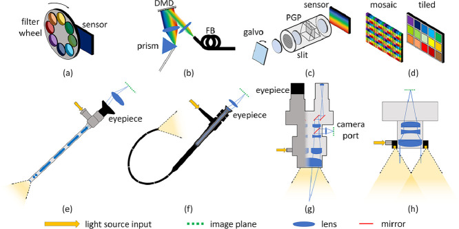
          Recent technological developments have resulted in the availability of miniaturised spectral imaging sensors capable of operating in the multi- (MSI) and hyperspectral imaging (HSI) regimes. Simultaneous advances in image-processing techniques and artificial intelligence (AI), especially in machine learning and deep learning, have made these data-rich modalities highly attractive as a means of extracting biological information non-destructively. Surgery in particular is poised to benefit from this, as spectrally-resolved tissue optical properties can offer enhanced contrast as well as diagnostic and guidance information during interventions. This is particularly relevant for procedures where inherent contrast is low under standard white light visualisation. This review summarises recent work in surgical spectral imaging (SSI) techniques, taken from Pubmed, Google Scholar and arXiv searches spanning the period 2013–2019. New hardware, optimised for use in both open and minimally-invasive surgery (MIS), is described, and recent commercial activity is summarised. Computational approaches to extract spectral information from conventional colour images are reviewed, as tip-mounted cameras become more commonplace in MIS. Model-based and machine learning methods of data analysis are discussed in addition to simulation, phantom and clinical validation experiments. A wide variety of surgical pilot studies are reported but it is apparent that further work is needed to quantify the clinical value of MSI/HSI. The current trend toward data-driven analysis emphasises the importance of widely-available, standardised spectral imaging datasets, which will aid understanding of variability across organs and patients, and drive clinical translation.

          Graphical abstract

          Related collections

          Most cited references183

          • Record: found
          • Abstract: found
          • Article: not found

          A survey on deep learning in medical image analysis

          Deep learning algorithms, in particular convolutional networks, have rapidly become a methodology of choice for analyzing medical images. This paper reviews the major deep learning concepts pertinent to medical image analysis and summarizes over 300 contributions to the field, most of which appeared in the last year. We survey the use of deep learning for image classification, object detection, segmentation, registration, and other tasks. Concise overviews are provided of studies per application area: neuro, retinal, pulmonary, digital pathology, breast, cardiac, abdominal, musculoskeletal. We end with a summary of the current state-of-the-art, a critical discussion of open challenges and directions for future research.
            Bookmark
            • Record: found
            • Abstract: found
            • Article: not found

            The Cancer Imaging Archive (TCIA): maintaining and operating a public information repository.

            The National Institutes of Health have placed significant emphasis on sharing of research data to support secondary research. Investigators have been encouraged to publish their clinical and imaging data as part of fulfilling their grant obligations. Realizing it was not sufficient to merely ask investigators to publish their collection of imaging and clinical data, the National Cancer Institute (NCI) created the open source National Biomedical Image Archive software package as a mechanism for centralized hosting of cancer related imaging. NCI has contracted with Washington University in Saint Louis to create The Cancer Imaging Archive (TCIA)-an open-source, open-access information resource to support research, development, and educational initiatives utilizing advanced medical imaging of cancer. In its first year of operation, TCIA accumulated 23 collections (3.3 million images). Operating and maintaining a high-availability image archive is a complex challenge involving varied archive-specific resources and driven by the needs of both image submitters and image consumers. Quality archives of any type (traditional library, PubMed, refereed journals) require management and customer service. This paper describes the management tasks and user support model for TCIA.
              Bookmark
              • Record: found
              • Abstract: found
              • Article: found
              Is Open Access

              Deep Convolutional Neural Networks for Computer-Aided Detection: CNN Architectures, Dataset Characteristics and Transfer Learning

              Remarkable progress has been made in image recognition, primarily due to the availability of large-scale annotated datasets and deep convolutional neural networks (CNNs). CNNs enable learning data-driven, highly representative, hierarchical image features from sufficient training data. However, obtaining datasets as comprehensively annotated as ImageNet in the medical imaging domain remains a challenge. There are currently three major techniques that successfully employ CNNs to medical image classification: training the CNN from scratch, using off-the-shelf pre-trained CNN features, and conducting unsupervised CNN pre-training with supervised fine-tuning. Another effective method is transfer learning, i.e., fine-tuning CNN models pre-trained from natural image dataset to medical image tasks. In this paper, we exploit three important, but previously understudied factors of employing deep convolutional neural networks to computer-aided detection problems. We first explore and evaluate different CNN architectures. The studied models contain 5 thousand to 160 million parameters, and vary in numbers of layers. We then evaluate the influence of dataset scale and spatial image context on performance. Finally, we examine when and why transfer learning from pre-trained ImageNet (via fine-tuning) can be useful. We study two specific computer-aided detection (CADe) problems, namely thoraco-abdominal lymph node (LN) detection and interstitial lung disease (ILD) classification. We achieve the state-of-the-art performance on the mediastinal LN detection, and report the first five-fold cross-validation classification results on predicting axial CT slices with ILD categories. Our extensive empirical evaluation, CNN model analysis and valuable insights can be extended to the design of high performance CAD systems for other medical imaging tasks.
                Bookmark

                Author and article information

                Contributors
                Journal
                Med Image Anal
                Med Image Anal
                Medical Image Analysis
                Elsevier
                1361-8415
                1361-8423
                1 July 2020
                July 2020
                : 63
                : 101699
                Affiliations
                [a ]Wellcome/EPSRC Centre for Interventional and Surgical Sciences (WEISS) , University College London , United Kingdom
                [b ]Centre for Medical Image Computing (CMIC), Department of Medical Physics and Biomedical Engineering, University College London, United Kingdom
                [c ]Centre for Medical Image Computing (CMIC), Department of Computer Science, University College London, United Kingdom
                [d ]German Cancer Research Centre (DKFZ), Heidelberg, Germany
                [e ]Hamlyn Centre for Robotic Surgery, Institute of Global Health Innovation, Imperial College London, United Kingdom
                [f ]Department of Surgery and Cancer, Imperial College London, United Kingdom
                Author notes
                [* ]Corresponding author at: WEISS, Charles Bell House, 43-45 Foley St., London, W1W 7TS, UK. n.clancy@ 123456ucl.ac.uk
                Article
                S1361-8415(20)30064-5 101699
                10.1016/j.media.2020.101699
                7903143
                32375102
                aae6f125-7834-4d0b-8439-64d0578b7986
                © 2020 The Authors. Published by Elsevier B.V.

                This is an open access article under the CC BY license (http://creativecommons.org/licenses/by/4.0/).

                History
                : 22 May 2019
                : 30 March 2020
                : 6 April 2020
                Categories
                Article

                Radiology & Imaging
                multispectral imaging,hyperspectral imaging,minimally-invasive surgery,computational imaging,ai, artificial intelligence,aotf, acousto-optic tuneable filter,cnn, convolutional neural network,ct, computed tomography,dmd, digital micromirror device,dpf, differential pathlength factor,emccd, electron-multiplying charge-coupled device,figs, fluorescence image-guided surgery,fwhm, full-width at half-maximum,gi, gastrointestinal,hsi, hyperspectral imaging,inn, invertible neural network,lctf, liquid crystal tuneable filter,led, light emitting diode,loocv, leave-one-out cross validation,mis, minimally-invasive surgery,mri, magnetic resonance imaging,msi, multispectral imaging,nbi, narrowband imaging,nir, near infrared,oem, original equipment manufacturer,rgb, red, green, blue,scmos, scientific complementary metal-oxide-semiconductor,sfdi, spatial frequency domain imaging,snr, signal-to-noise ratio,ssi, surgical spectral imaging,svm, support vector machine,vof, variable optical filter

                Comments

                Comment on this article