7
views
0
recommends
+1 Recommend
0 collections
    0
    shares
      • Record: found
      • Abstract: found
      • Article: found
      Is Open Access

      Friend and foe: β-cell Ca 2+ signaling and the development of diabetes

      review-article

      Read this article at

      Bookmark
          There is no author summary for this article yet. Authors can add summaries to their articles on ScienceOpen to make them more accessible to a non-specialist audience.

          Abstract

          Background

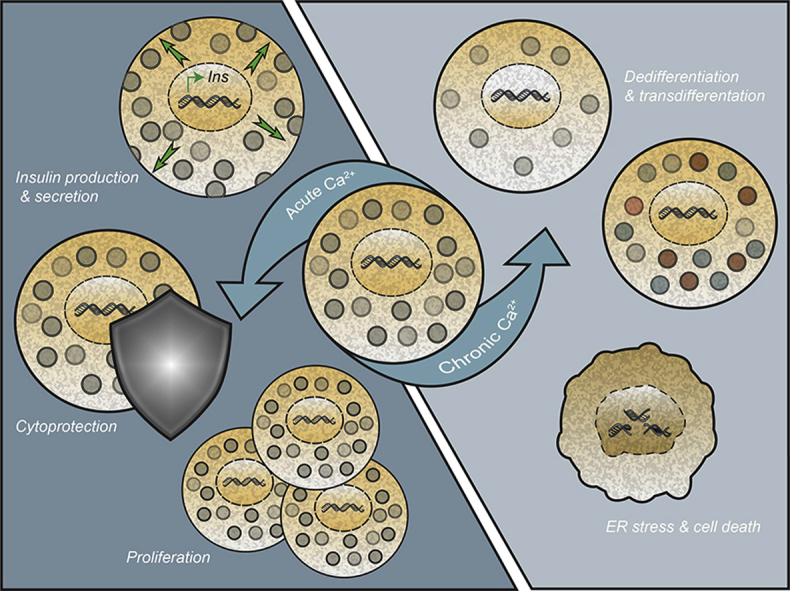
          The divalent cation Calcium (Ca 2+) regulates a wide range of processes in disparate cell types. Within insulin-producing β-cells, increases in cytosolic Ca 2+ directly stimulate insulin vesicle exocytosis, but also initiate multiple signaling pathways. Mediated through activation of downstream kinases and transcription factors, Ca 2+-regulated signaling pathways leverage substantial influence on a number of critical cellular processes within the β-cell. Additionally, there is evidence that prolonged activation of these same pathways is detrimental to β-cell health and may contribute to Type 2 Diabetes pathogenesis.

          Scope of review

          This review aims to briefly highlight canonical Ca 2+ signaling pathways in β-cells and how β-cells regulate the movement of Ca 2+ across numerous organelles and microdomains. As a main focus, this review synthesizes experimental data from in vitro and in vivo models on both the beneficial and detrimental effects of Ca 2+ signaling pathways for β-cell function and health.

          Major conclusions

          Acute increases in intracellular Ca 2+ stimulate a number of signaling cascades, resulting in (de-)phosphorylation events and activation of downstream transcription factors. The short-term stimulation of these Ca 2+ signaling pathways promotes numerous cellular processes critical to β-cell function, including increased viability, replication, and insulin production and secretion. Conversely, chronic stimulation of Ca 2+ signaling pathways increases β-cell ER stress and results in the loss of β-cell differentiation status. Together, decades of study demonstrate that Ca 2+ movement is tightly regulated within the β-cell, which is at least partially due to its dual roles as a potent signaling molecule.

          Graphical abstract

          Related collections

          Most cited references133

          • Record: found
          • Abstract: found
          • Article: not found

          NCLX is an essential component of mitochondrial Na+/Ca2+ exchange.

          Mitochondrial Ca(2+) efflux is linked to numerous cellular activities and pathophysiological processes. Although it is established that an Na(+)-dependent mechanism mediates mitochondrial Ca(2+) efflux, the molecular identity of this transporter has remained elusive. Here we show that the Na(+)/Ca(2+) exchanger NCLX is enriched in mitochondria, where it is localized to the cristae. Employing Ca(2+) and Na(+) fluorescent imaging, we demonstrate that mitochondrial Na(+)-dependent Ca(2+) efflux is enhanced upon overexpression of NCLX, is reduced by silencing of NCLX expression by siRNA, and is fully rescued by the concomitant expression of heterologous NCLX. NCLX-mediated mitochondrial Ca(2+) transport was inhibited, moreover, by CGP-37157 and exhibited Li(+) dependence, both hallmarks of mitochondrial Na(+)-dependent Ca(2+) efflux. Finally, NCLX-mediated mitochondrial Ca(2+) exchange is blocked in cells expressing a catalytically inactive NCLX mutant. Taken together, our results converge to the conclusion that NCLX is the long-sought mitochondrial Na(+)/Ca(2+) exchanger.
            Bookmark
            • Record: found
            • Abstract: found
            • Article: not found

            NAADP mobilizes calcium from acidic organelles through two-pore channels

            Ca2+ mobilization from intracellular stores represents an important cell signaling process 1 which is regulated, in mammalian cells, by inositol 1,4,5-trisphosphate (InsP3), cyclic ADP ribose (cADPR) and nicotinic acid adenine dinucleotide phosphate (NAADP). InsP3 and cADPR release Ca2+ from sarco / endoplasmic reticulum (S/ER) stores through activation of InsP3 and ryanodine receptors (InsP3Rs and RyRs). By contrast, the nature of the intracellular stores targeted by NAADP and molecular identity of the NAADP receptors remain controversial 1,2, although evidence indicates that NAADP mobilizes Ca2+ from lysosome-related acidic compartments 3,4. Here we show that two-pore channels (TPCs) comprise a family of NAADP receptors, with TPC1 and TPC3 being expressed on endosomal and TPC2 on lysosomal membranes. Membranes enriched with TPC2 exhibit high affinity NAADP binding and TPC2 underpins NAADP-induced Ca2+ release from lysosome-related stores that is subsequently amplified by Ca2+-induced Ca2+ release via InsP3Rs. Responses to NAADP were abolished by disrupting the lysosomal proton gradient and by ablating TPC2 expression, but only attenuated by depleting ER Ca2+ stores or blocking InsP3Rs. Thus, TPCs form NAADP receptors that release Ca2+ from acidic organelles, which can trigger additional Ca2+ signals via S/ER. TPCs therefore provide new insights into the regulation and organization of Ca2+ signals in animal cells and will advance our understanding of the physiological role of NAADP.
              Bookmark
              • Record: found
              • Abstract: found
              • Article: not found

              Metabolic signaling in fuel-induced insulin secretion.

              The pancreatic islet β cell senses circulating levels of calorigenic nutrients to secrete insulin according to the needs of the organism. Altered insulin secretion is linked to various disorders such as diabetes, hypoglycemic states, and cardiometabolic diseases. Fuel stimuli, including glucose, free fatty acids, and amino acids, promote insulin granule exocytosis primarily via their metabolism in β cells and the production of key signaling metabolites. This paper reviews our current knowledge of the pathways involved in both positive and negative metabolic signaling for insulin secretion and assesses the role of established and candidate metabolic coupling factors, keeping recent developments in focus. Copyright © 2013 Elsevier Inc. All rights reserved.
                Bookmark

                Author and article information

                Contributors
                Journal
                Mol Metab
                Mol Metab
                Molecular Metabolism
                Elsevier
                2212-8778
                24 December 2018
                March 2019
                24 December 2018
                : 21
                : 1-12
                Affiliations
                [1 ]Diabetes Research Group, BC Children's Hospital Research Institute, Vancouver, British Columbia, Canada
                [2 ]Department of Surgery, University of British Columbia, Vancouver, British Columbia, Canada
                [3 ]Department of Cellular and Physiological Sciences, University of British Columbia, Vancouver, British Columbia, Canada
                [4 ]Department of Internal Medicine, University of Michigan, Ann Arbor, MI, USA
                Author notes
                []Corresponding author. 950 28th Ave W, Vancouver, BC, V5Z 4H4, Canada. Fax: +604 875 2373. francis.lynn@ 123456ubc.ca @ 123456nictitate
                Article
                S2212-8778(18)30896-2
                10.1016/j.molmet.2018.12.007
                6407368
                30630689
                a2543bb3-f8bb-4a68-8406-c64bb4ee945a
                © 2018 The Authors

                This is an open access article under the CC BY license (http://creativecommons.org/licenses/by/4.0/).

                History
                : 6 September 2018
                : 3 December 2018
                : 19 December 2018
                Categories
                Article

                β-cells,diabetes,ca2+,creb,nfat,calmodulin,calcineurin,camk
                β-cells, diabetes, ca2+, creb, nfat, calmodulin, calcineurin, camk

                Comments

                Comment on this article