104
views
0
recommends
+1 Recommend
1 collections
    0
    shares
      • Record: found
      • Abstract: found
      • Article: found
      Is Open Access

      The Major Risk Factors for Alzheimer’s Disease: Age, Sex, and Genes Modulate the Microglia Response to Aβ Plaques

      research-article

      Read this article at

      ScienceOpenPublisherPMC
      Bookmark
          There is no author summary for this article yet. Authors can add summaries to their articles on ScienceOpen to make them more accessible to a non-specialist audience.

          SUMMARY

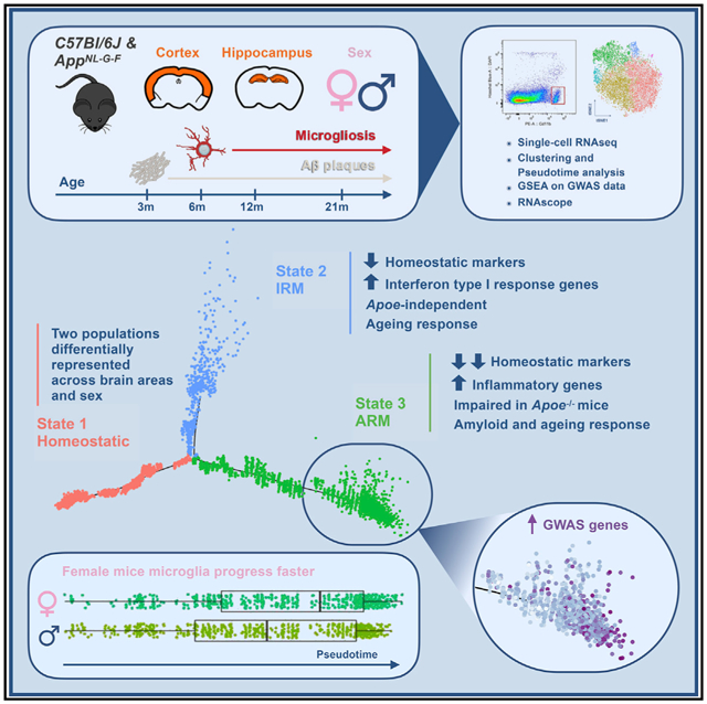
          Gene expression profiles of more than 10,000 individual microglial cells isolated from cortex and hippocampus of male and female App NL-G-F mice over time demonstrate that progressive amyloid-b accumulation accelerates two main activated microglia states that are also present during normal aging. Activated response microglia (ARMs) are composed of specialized subgroups overexpressing MHC type II and putative tissue repair genes ( Dkk2, Gpnmb, and Spp1) and are strongly enriched with Alzheimer’s disease (AD) risk genes. Microglia from female mice progress faster in this activation trajectory. Similar activated states are also found in a second AD model and in human brain. Apoe, the major genetic risk factor for AD, regulates the ARMs but not the interferon response microglia (IRMs). Thus, the ARMs response is the converging point for aging, sex, and genetic AD risk factors.

          Graphical Abstract

          In Brief

          Sala Frigerio et al. show how microglia respond to amyloid-b, the Alzheimer’s disease (AD)-causing factor. Their major response, the ARMs response, is enriched for AD risk genes, is abolished by Apoe deletion, develops faster in female mice, and is also part of normal aging. Thus, major AD risk factors converge on microglia.

          Related collections

          Most cited references27

          • Record: found
          • Abstract: found
          • Article: not found

          Gene dose of apolipoprotein E type 4 allele and the risk of Alzheimer's disease in late onset families.

          The apolipoprotein E type 4 allele (APOE-epsilon 4) is genetically associated with the common late onset familial and sporadic forms of Alzheimer's disease (AD). Risk for AD increased from 20% to 90% and mean age at onset decreased from 84 to 68 years with increasing number of APOE-epsilon 4 alleles in 42 families with late onset AD. Thus APOE-epsilon 4 gene dose is a major risk factor for late onset AD and, in these families, homozygosity for APOE-epsilon 4 was virtually sufficient to cause AD by age 80.
            Bookmark
            • Record: found
            • Abstract: found
            • Article: not found

            TREM2 Binds to Apolipoproteins, Including APOE and CLU/APOJ, and Thereby Facilitates Uptake of Amyloid-Beta by Microglia.

            Genetic variants of TREM2, a protein expressed selectively by microglia in the brain, are associated with Alzheimer's disease (AD). Starting from an unbiased protein microarray screen, we identified a set of lipoprotein particles (including LDL) and apolipoproteins (including CLU/APOJ and APOE) as ligands of TREM2. Binding of these ligands by TREM2 was abolished or reduced by disease-associated mutations. Overexpression of wild-type TREM2 was sufficient to enhance uptake of LDL, CLU, and APOE in heterologous cells, whereas TREM2 disease variants were impaired in this activity. Trem2 knockout microglia showed reduced internalization of LDL and CLU. β-amyloid (Aβ) binds to lipoproteins and this complex is efficiently taken up by microglia in a TREM2-dependent fashion. Uptake of Aβ-lipoprotein complexes was reduced in macrophages from human subjects carrying a TREM2 AD variant. These data link three genetic risk factors for AD and reveal a possible mechanism by which mutant TREM2 increases risk of AD. VIDEO ABSTRACT.
              Bookmark
              • Record: found
              • Abstract: found
              • Article: not found

              Microglial signatures and their role in health and disease

              Microglia are the primary innate immune cells in the CNS. In the healthy brain, they exhibit a unique molecular homeostatic ‘signature’, consisting of a specific transcriptional profile and surface protein expression pattern, which differs from that of tissue macrophages. In recent years, there have been a number of important advances in our understanding of the molecular signatures of homeostatic microglia and disease-associated microglia that have provided insight into how these cells are regulated in health and disease and how they contribute to the maintenance of the neural environment.
                Bookmark

                Author and article information

                Journal
                101573691
                39703
                Cell Rep
                Cell Rep
                Cell reports
                2211-1247
                26 May 2020
                23 April 2019
                07 July 2020
                : 27
                : 4
                : 1293-1306.e6
                Affiliations
                [1 ]VIB Centre for Brain Disease Research, Leuven, Belgium
                [2 ]University of Leuven, Department of Neurosciences and Leuven Brain Institute, Leuven, Belgium
                [3 ]UK Dementia Research Institute, University College London, London, UK
                [4 ]Foundational Neuroscience Center, AbbVie, Inc., Cambridge, MA, USA
                [5 ]Department of Neurology, MassGeneral Institute for Neurodegenerative Disease, Massachusetts General Hospital, Harvard Medical School, Charlestown, MA, USA
                [6 ]Laboratory for Proteolytic Neuroscience, RIKEN Brain Science Institute, Wako-shi, Saitama, Japan
                [7 ]Centre for Biological Sciences, University of Southampton, Southampton, UK
                [8 ]Lead Contact
                Author notes

                AUTHOR CONTRIBUTIONS

                C.S.F. and B.D.S. conceptualized and designed the study and the experiments. C.S.F. optimized methods with L.W. and analyzed and interpreted single-cell sequencing and imaging data. L.W. isolated microglia, prepared single-cell RNA-sequencing libraries, performed in situ RNA hybridization and immunofluorescent staining, and analyzed and interpreted in situ data.I.V. and I.S. performed in situ RNA hybridization and immunofluorescent staining and analyzed and interpreted imaging data. N.F., N.T., W.-T.C., M.E.W., G.S., and M.F. analyzed and interpreted single-cell RNA-sequencing data. E.H. performed amyloid burden analysis. S.D. analyzed human RNA-sequencing data. R.M., T.M., E.K., B.H., and V.H.P. interpreted data. T.S. and B.H. contributed animals. B.D.S. procured funding and interpreted data. The manuscript was written by C.S.F. and B.D.S., with input from all co-authors. All co-authors read and approved the final version of the manuscript.

                Article
                NIHMS1596077
                10.1016/j.celrep.2019.03.099
                7340153
                31018141
                956ffb9b-5483-463c-8b28-804a3801c33c

                This is an open access article under the CC BY-NC-ND license ( http://creativecommons.org/licenses/by-nc-nd/4.0/).

                History
                Categories
                Article

                Cell biology
                Cell biology

                Comments

                Comment on this article