6
views
0
recommends
+1 Recommend
0 collections
    0
    shares
      • Record: found
      • Abstract: found
      • Article: found
      Is Open Access

      Possible mechanisms of lycopene amelioration of learning and memory impairment in rats with vascular dementia

      research-article

      Read this article at

      Bookmark
          There is no author summary for this article yet. Authors can add summaries to their articles on ScienceOpen to make them more accessible to a non-specialist audience.

          Abstract

          Abstract

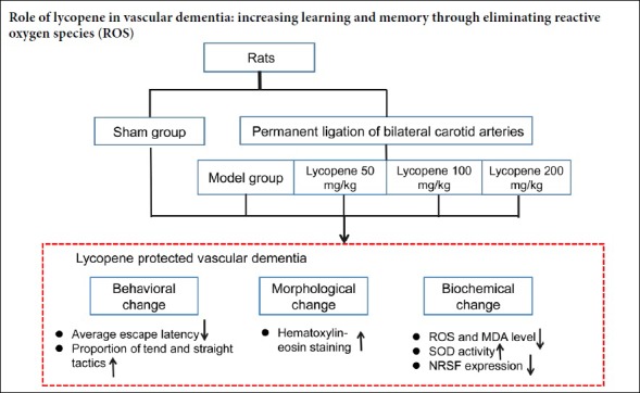
          Oxidative stress is involved in the pathogenesis of vascular dementia. Studies have shown that lycopene can significantly inhibit oxidative stress; therefore, we hypothesized that lycopene can reduce the level of oxidative stress in vascular dementia. A vascular dementia model was established by permanent bilateral ligation of common carotid arteries. The dosage groups were treated with lycopene (50, 100 and 200 mg/kg) every other day for 2 months. Rats without bilateral carotid artery ligation were prepared as a sham group. To test the ability of learning and memory, the Morris water maze was used to detect the average escape latency and the change of search strategy. Hematoxylin-eosin staining was used to observe changes of hippocampal neurons. The levels of oxidative stress factors, superoxide dismutase and malondialdehyde, were measured in the hippocampus by biochemical detection. The levels of reactive oxygen species in the hippocampus were observed by dihydroethidium staining. The distribution and expression of oxidative stress related protein, neuron-restrictive silencer factor, in hippocampal neurons were detected by immunofluorescence histochemistry and western blot assays. After 2 months of drug administration, (1) in the model group, the average escape latency was longer than that of the sham group, and the proportion of straight and tend tactics was lower than that of the sham group, and the hippocampal neurons were irregularly arranged and the cytoplasm was hyperchromatic. (2) The levels of reactive oxygen species and malondialdehyde in the hippocampus of the model group rats were increased, and the activity of superoxide dismutase was decreased. (3) Lycopene (50, 100 and 200 mg/kg) intervention improved the above changes, and the lycopene 100 mg/kg group showed the most significant improvement effect. (4) Neuron-restrictive silencer factor expression in the hippocampus was lower in the sham group and the lycopene 100 mg/kg group than in the model group. (5) The above data indicate that lycopene 100 mg/kg could protect against the learning-memory ability impairment of vascular dementia rats. The protective mechanism was achieved by inhibiting oxidative stress in the hippocampus. The experiment was approved by the Animal Ethics Committee of Fujian Medical University, China (approval No. 2014-025) in June 2014.

          Related collections

          Most cited references49

          • Record: found
          • Abstract: found
          • Article: not found

          REST: a mammalian silencer protein that restricts sodium channel gene expression to neurons.

          Expression of the type II voltage-dependent sodium channel gene is restricted to neurons by a silencer element active in nonneuronal cells. We have cloned cDNA coding for a transcription factor (REST) that binds to this silencer element. Expression of a recombinant REST protein confers the ability to silence type II reporter genes in neuronal cell types lacking the native REST protein, whereas expression of a dominant negative form of REST in nonneuronal cells relieves silencing mediated by the native protein. REST transcripts in developing mouse embryos are detected ubiquitously outside of the nervous system. We propose that expression of the type II sodium channel gene in neurons reflects a default pathway that is blocked in nonneuronal cells by the presence of REST.
            Bookmark
            • Record: found
            • Abstract: found
            • Article: not found

            Role of brain-derived neurotrophic factor in Huntington's disease.

            Neurotrophic factors are essential contributors to the survival of peripheral and central nervous system (CNS) neurons, and demonstration of their reduced availability in diseased brains indicates that they play a role in various neurological disorders. This paper will concentrate on the role of brain-derived neurotrophic factor (BDNF) in the survival and activity of the neurons that die in Huntington's disease (HD) by reviewing the evidence indicating that it involves profound changes in BDNF levels and that attempts to restore these levels are therapeutically interesting. BDNF is a small dimeric protein that is widely expressed in adult mammalian brain and has been shown to promote the survival of all major neuronal types affected in Alzheimer's disease (AD) and Parkinson's disease (PD). Furthermore, cortical BDNF production is required for the correct activity of the corticostriatal synapse and the survival of the GABA-ergic medium-sized spiny striatal neurons that die in HD. We will highlight the available data concerning changes in BDNF levels in HD cells, mice and human postmortem samples, describe the molecular evidence underlying this alteration, and review the data concerning the impact of the experimental manipulation of BDNF levels on HD progression. Such studies have revealed a major loss of BDNF protein in the striatum of HD patients which may contribute to the clinical manifestations of the disease. They have also opened up a molecular window into the underlying pathogenic mechanism and new therapeutic perspectives by raising the possibility that one of the mechanisms triggering the reduction in BDNF in HD may also affect the activity of many other neuronal proteins.
              Bookmark
              • Record: found
              • Abstract: found
              • Article: not found

              Genome-wide analysis of repressor element 1 silencing transcription factor/neuron-restrictive silencing factor (REST/NRSF) target genes.

              The completion of whole genome sequencing projects has provided the genetic instructions of life. However, whereas the identification of gene coding regions has progressed, the mapping of transcriptional regulatory motifs has moved more slowly. To understand how distinct expression profiles can be established and maintained, a greater understanding of these sequences and their trans-acting factors is required. Herein we have used a combined in silico and biochemical approach to identify binding sites [repressor element 1/neuron-restrictive silencer element (RE1/NRSE)] and potential target genes of RE1 silencing transcription factor/neuron-restrictive silencing factor (REST/NRSF) within the human, mouse, and Fugu rubripes genomes. We have used this genome-wide analysis to identify 1,892 human, 1,894 mouse, and 554 Fugu RE1/NRSEs and present their location and gene linkages in a searchable database. Furthermore, we identified an in vivo hierarchy in which distinct subsets of RE1/NRSEs interact with endogenous levels of REST/NRSF, whereas others function as bona fide transcriptional control elements only in the presence of elevated levels of REST/NRSF. These data show that individual RE1/NRSE sites interact differentially with REST/NRSF within a particular cell type. This combined bioinformatic and biochemical approach serves to illustrate the selective manner in which a transcription factor interacts with its potential binding sites and regulates target genes. In addition, this approach provides a unique whole-genome map for a given transcription factor-binding site implicated in establishing specific patterns of neuronal gene expression.
                Bookmark

                Author and article information

                Journal
                Neural Regen Res
                Neural Regen Res
                NRR
                Neural Regeneration Research
                Wolters Kluwer - Medknow (India )
                1673-5374
                1876-7958
                February 2020
                24 September 2019
                : 15
                : 2
                : 332-341
                Affiliations
                [1 ]Department of Human Anatomy, Histology and Embryology, School of Basic Medical Sciences, Fujian Medical University, Fuzhou, Fujian Province, China
                [2 ]Department of Pharmacy, Zhejiang Pharmaceutical College, Ningbo, Zhejiang Province, China
                [3 ]Key Laboratory of Brain Aging and Neurodegenerative Diseases, School of Basic Medical Sciences, Fujian Medical University, Fuzhou, Fujian Province, China
                Author notes
                [* ] Correspondence to: Xiao-Zhen Zhao, zxz8338@ 123456163.com .

                Author contributions: Study concept and design, data collection, analysis and interpretation: XZZ; manuscript writing, critical revision of the manuscript for intellectual content: NWZ, XLY, RL; statistical expertise, fundraising administrative, technical or material support, and supervision: XLF, SJC, YMZ. All authors approved the final version of the paper. .

                Author information
                http://orcid.org/0000-0002-5546-9151
                Article
                NRR-15-332
                10.4103/1673-5374.265565
                6905346
                31552907
                1d5e55db-0c1e-4e6b-ac04-3b1a18dcf9bb
                Copyright: © Neural Regeneration Research

                This is an open access journal, and articles are distributed under the terms of the Creative Commons Attribution-NonCommercial-ShareAlike 4.0 License, which allows others to remix, tweak, and build upon the work non-commercially, as long as appropriate credit is given and the new creations are licensed under the identical terms.

                History
                : 28 September 2018
                : 23 May 2019
                Categories
                Research Article

                bilateral carotid artery ligation,lycopene,hippocampus,learning and memory,malondialdehyde,neuron,neuron-restrictive silencer factor,reactive oxygen species,superoxide dismutase

                Comments

                Comment on this article